CellPAINT: Digital Illustration from Atoms to Cells
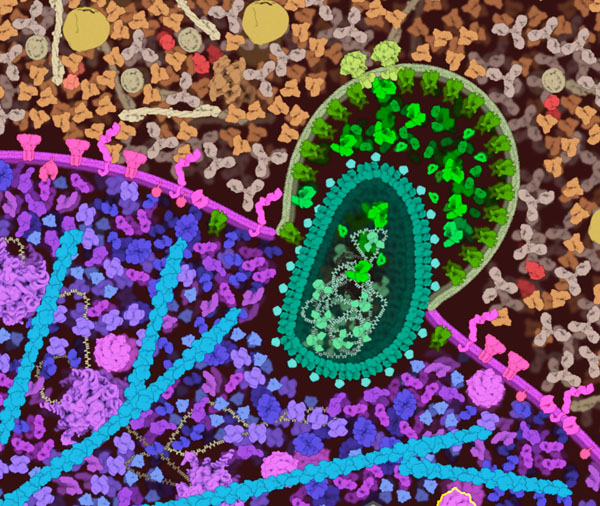
With CellPAINT, you can create dynamic illustrations of HIV in blood plasma, along with the surface of a T-cell. CellPAINT is being developed as part of an effort to model the molecular structure of whole living cells. It will be used in two ways: to allow researchers to sketch out scenes based on their research, which may then be used to guide the construction of detailed 3-D models of the scene, and for use in education and outreach to integrate and present the results of scientific research.
The program is being developed by Adam Gardner, Ludovic Autin, Arthur Olson and David Goodsell, with support from the US National Institutes of Health.

The Science in CellPAINT
CellPAINT pictures are based on experimental results from many different laboratories. The shape of the HIV envelope and capsid may be based on an electron micrograph, as displayed in the tool panel. The size and shape of each molecule is based on atomic structures determined by x-ray crystallography, NMR spectroscopy and electron microscopy. These molecules also interact in complex ways. CellPAINT currently shows some of these interactions, such as the interaction of cyclophilin with capsid and the interaction of nucleocapsid with RNA. Other user-defined interactions will be possible in future versions of the program.
CellPAINT-VR
CellPAINT-VR is a virtual reality implementation of CellPAINT. Like CellPAINT it is an interactive visualization tool in which molecular structures can be positioned in a workspace to create mesoscale scenes of biological macromolecules in complex cellular environments. Unlike CellPAINT it is an immersive 3d stereoscopic implementation that runs on high-end head mounted displays, including those from Oculus, HTC Vive, and Windows Mixed Reality. The current implementation runs only on the Microsoft Windows10 operating system with a sufficiently powerful NVidia Graphics Processing Unit.
Inspired by CellPAINT it presents palates of molecular structures that can be selected and interactively placed into a scene. Membrane-bound compartments can be created from isosurfaces of interactively manipulated spherical functions. The 3D molecular models can be placed into open space, inside compartments or on membrane surfaces depending on functional type. Fibrous molecules like DNA and RNA can be interactively drawn in three dimensions. Molecular representation can switch between coarse-grain polyhedral surfaces and full atomic CPK renderings as produced by CellView/CellPACKgpu. Tool functions include painting/placing/spraying, erasing, moving/rotating, pinning, querying/coloring, and creating complexes from multiple molecules. Like CellPAINT one can simulate diffusion using Brownian motion. Navigation within the space, and the creation and manipulation of the objects in the scene are accomplished with two 6 degree of freedom hand controllers. In this sense CellPAINT-VR has morphed from the original painting metaphor into a sculptural or world-building one.
CellPAINT-Exo
CellPAINT-Exo is our first experiment with importing hand-drawn molecules into CellPAINT. As part of an internship in the lab, Julia Jiminez created a painting of exosomes (small vesicles released by many types of cells). She then created individual images of each of the molecules, and these were added to a custom version of CellPAINT.
Tell us what you think!
CellPAINT is currently in active development, and we are looking for testers. The software still has a few idiosyncracies, so please be prepared to restart the programs if you encounter problems. We are looking for feedback on several general questions:
Were you able to create the scenes that you wanted to make?
Will this type of program be useful in your own work?
Are there any features that you would like to see added?
Please send your impressions to David Goodsell at goodsell@scripps.edu.
You can download CellPAINT, CellPAINT-VR and CellPaint-Exo at SourceForge.
We also have a WebGL prototype–give it a try in your web browser!
