Scientist/Artist David S. Goodsell uses watercolor and molecular graphics to explore the cellular mesoscale
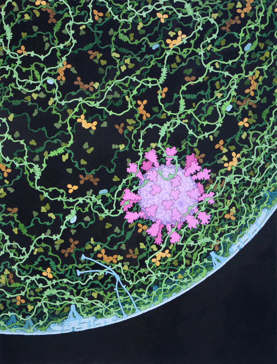
Respiratory Droplet
2020, Watercolor and Ink on Paper
This painting shows a cross section through a small respiratory droplet, like the ones that are thought to transmit SARS-CoV-2. The virus is shown in magenta, and the droplet is also filled with molecules that are present in the respiratory tract, including mucins (green), pulmonary surfactant proteins and lipids (blue), and antibodies (tan).
A full-size file is available under creative commons at the RCSB Protein Data Bank at doi: 10.2210/rcsb_pdb/goodsell-gallery-024

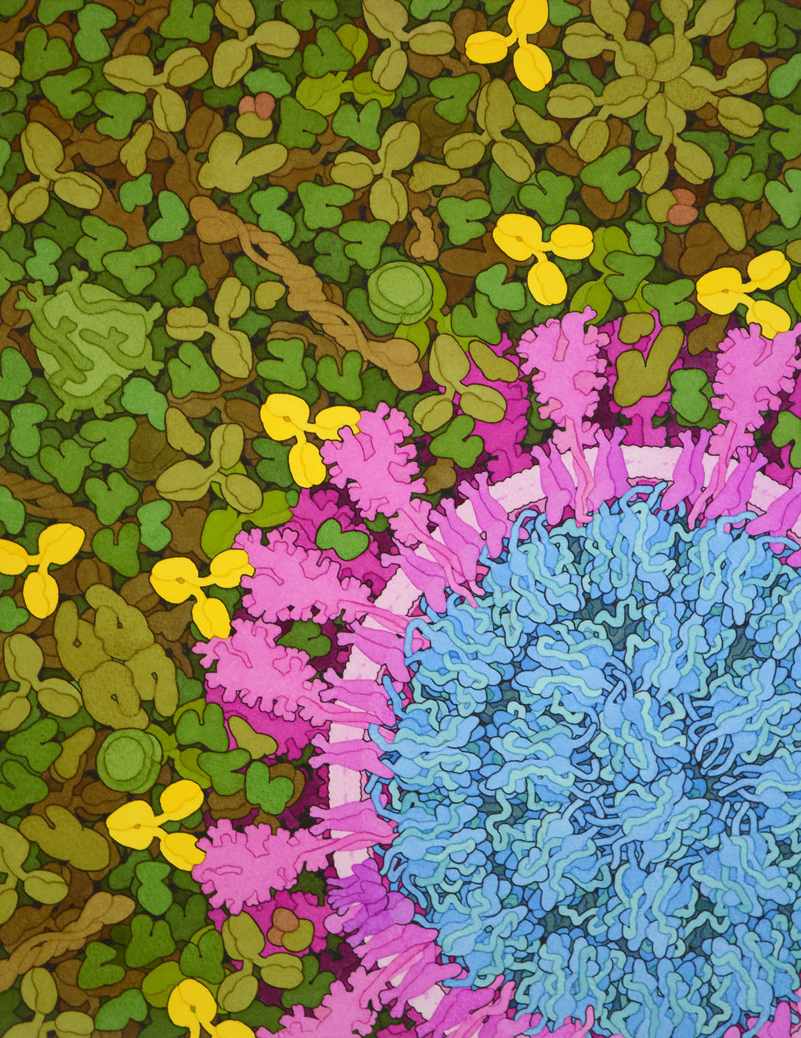
SARS-CoV-2 and Neutralizing Antibodies
2020, Watercolor and Ink on Paper
This painting shows a cross section through SARS-CoV-2 surrounded by blood plasma, with neutralizing antibodies in bright yellow. The painting was commissioned for the cover of a special COVID-19 issue of Nature, presented 20 August 2020.
It incorporates information from two cryoelectron microscopy studies that explore the shape and distribution of spikes and the nucleoprotein:
Yao H et al. (2020) Molecular architecture of the SARS-CoV-2 virus. bioRxiv preprint DOI: 10.1101/2020.07.08.192104
Ke Z et al. (2020) Structures, conformations and distributions of SARS-CoV-2 spike protein trimers on intact virions. bioRxiv preprint DOI: 10.1101/2020.06.27.174979
A full-size file is available under creative commons at the RCSB Protein Data Bank at doi: 10.2210/rcsb_pdb/goodsell-gallery-025

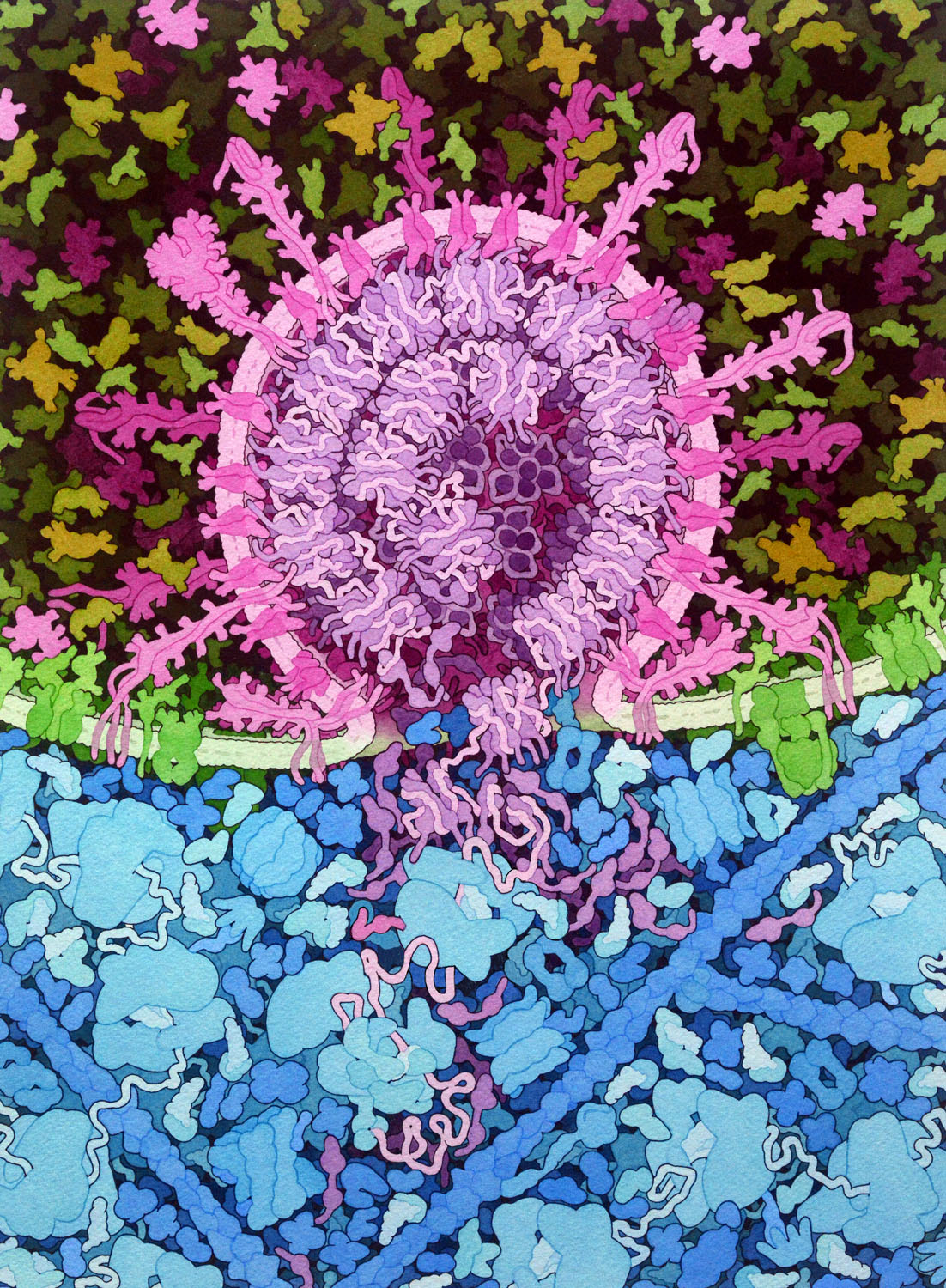
SARS-CoV-2 Fusion
2020, Watercolor and Ink on Paper
This painting depicts the fusion of SARS-CoV-2 (magenta) with an endosomal membrane (green), releasing the viral RNA genome into the cell cytoplasm (blue), where it is beginning to be translated by cellular ribosomes to create viral polyproteins. The painting includes speculative elements that are designed highlight the process, most notably, multiple states of the viral spike protein are shown.
A full-size file is available under creative commons at the RCSB Protein Data Bank at doi: 10.2210/rcsb_pdb/goodsell-gallery-026

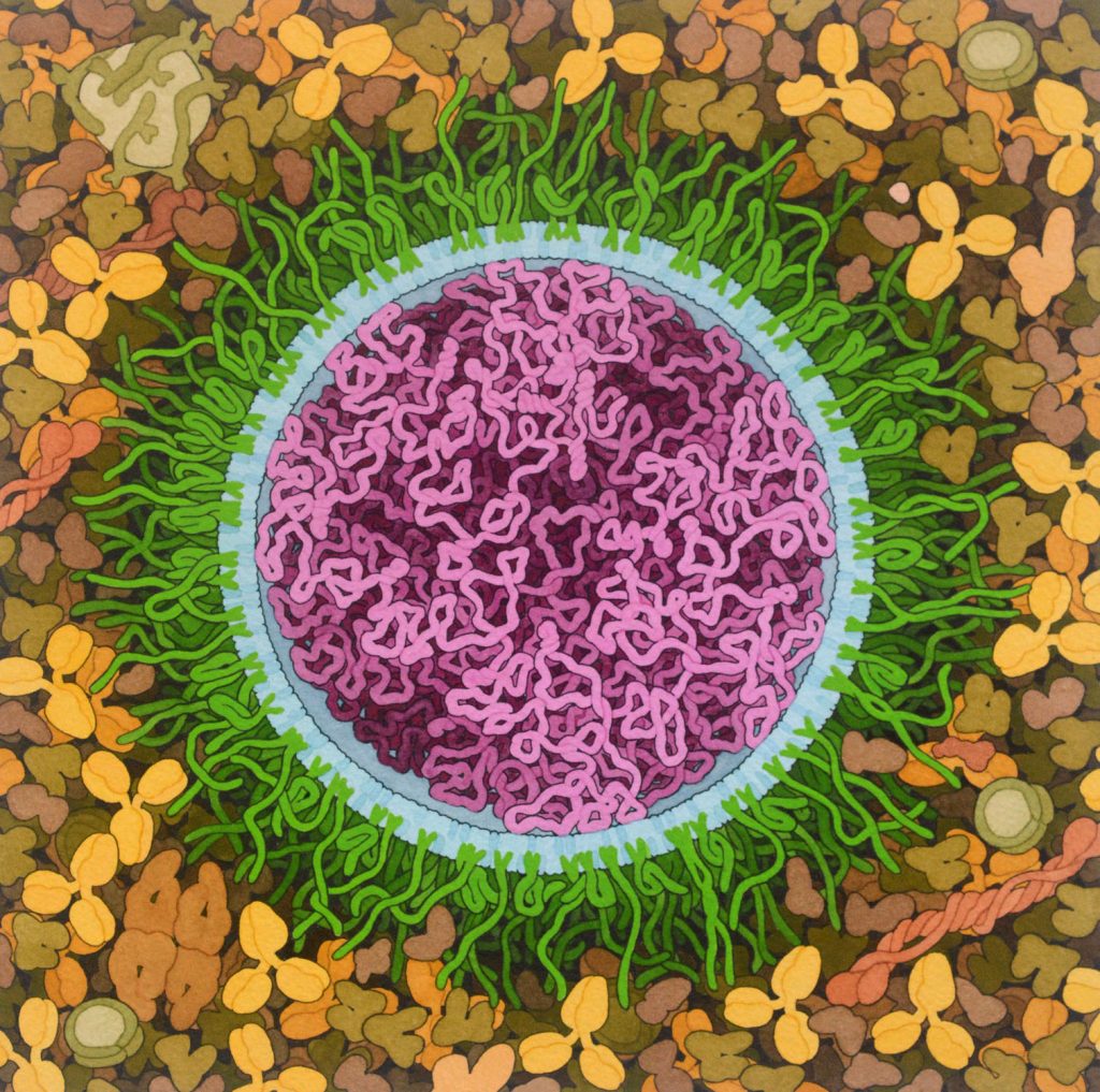
mRNA Vaccine
2020, Watercolor and Ink on Paper
Messenger RNA (mRNA) vaccines developed for the COVID-19 pandemic are composed of long strands of RNA (magenta) that encode the SARS-CoV-2 spike surface glycoprotein enclosed in lipids (blue) that deliver the RNA into cells. Several different types of lipids are used, including familar lipids, cholesterol, ionizable lipids that interact with RNA, and lipids connected to polyethylene glycol chains (green) that help shield the vaccine from the immune system, lengthening its lifetime following administration. In this idealized illustration, all of the lipids are arranged in a simple circular bilayer that surrounds the mRNA and the PEG strands have both extended and folded conformations. In reality, the structure may be less regular. Learn more about how this vaccine works in Resources to Fight the COVID-19 Pandemic at the RCSB Protein Data Bank.
A full-size file is available under creative commons at the RCSB Protein Data Bank at doi: 10.2210/rcsb_pdb/goodsell-gallery-027
